March 17 – May 10, 2009
The Benton’s contribution to the University-wide “Year of Science 2009” celebration is an exhibition that chronicles the history of medical illustration through a selection of prints, drawings, computer graphics and animation from the 16th century to the present. Each piece articulates a unique union of art, anatomy and medicine, the works together reflecting the ways that union has evolved over the centuries and continues to thrive in an era of digital photography and 3-D imaging.
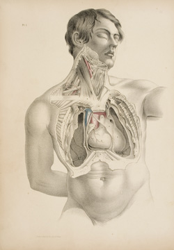
The works in Anatomically Correct come out of a pictorial tradition that was set in motion by the Belgian anatomist Andreas Vesalius (1514–1564). He conducted his dissections firsthand, breaking away from the longstanding authority of classical texts by relying on direct observation. He similarly reformed scientific illustration by insisting upon anatomical accuracy and precision for the plates in his pivotal work, De Humani Corporis Fabrica (On the Fabric of the Human Body) of 1543. These illustrations by the artist John Van Calcar divided the field of scientific illustration into “pre-Vesalian” and “post-Vesalian” periods. With Vesalius’s brilliant integration of image and text, illustration began to serve a crucial function in the communication of scientific information, initiating the role of scientific illustration as a record of the progress of science in general.
Along with Vesalius’s contributions, Anatomically Correct highlights significant post-Vesalian developments that carried his vision into the present. Illustrations from William Hunter’s The Anatomy of the Human Gravid Uterus (1774) played a vital role in establishing obstetrics as a field of medicine rather than a practice of midwives. A major advancement in print technology is represented by color lithography in Jean Marc Bourgery’s Atlas of Anatomy (1831–1854). A shift in the role of illustration from works of art in themselves to didactic tools can be seen in Henry Gray’s Anatomy: Descriptive and Surgical (1858), and the increased role of the computer in contemporary illustration is evidenced in the 3-D animation of the Connecticut-based XVIVO studio.
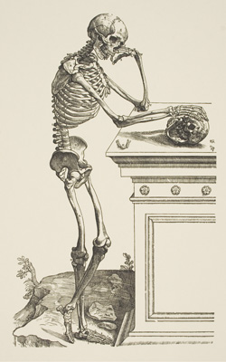
In a discipline we associate with objectivity and empiricism, the works in the exhibition consistently reveal themselves to be products of their respective social climates. Ideological and social conventions inevitably come through in the illustrations as anatomists and artist catered to their audience’s desire to see the body represented morally, socially, theologically.
Anatomically Correct commemorates the 200th anniversary of Charles Darwin’s birth. The exhibition is particularly relevant to this moment of celebration as a reflection on the advancement in the field of anatomy as a basis for the theory of evolution and as an acknowledgement of the role of art as critical to the way these advancements were visualized.
Exhibition curated by Eve Perry, M.A. candidate, Art History, 2009.